Embark on a journey into the depths of human physiology with our “Nervous System Flow Chart Worksheet.” This meticulously crafted resource unravels the complexities of the nervous system, providing a comprehensive understanding of its structure, functions, and intricate pathways.
Through engaging text, interactive flowcharts, and insightful explanations, this worksheet serves as an indispensable tool for students, educators, and healthcare professionals seeking to delve into the fascinating world of neural communication.
Nervous System Structure and Function

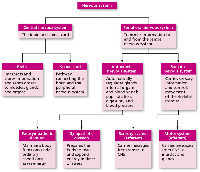
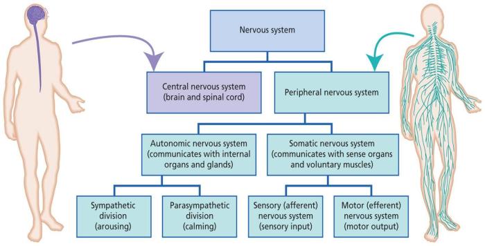
The nervous system is a complex network of cells, tissues, and organs that work together to control all bodily functions. It is divided into two main components: the central nervous system (CNS) and the peripheral nervous system (PNS).
The CNS consists of the brain and spinal cord. The brain is the center of the nervous system and controls all bodily functions, including thought, emotion, movement, and memory. The spinal cord is a long, thin bundle of nerves that runs from the brain down the back.
It carries messages between the brain and the rest of the body.
The PNS consists of all the nerves that connect the CNS to the rest of the body. These nerves are divided into two types: sensory nerves and motor nerves. Sensory nerves carry messages from the body to the brain, while motor nerves carry messages from the brain to the body.
Neural Communication
Neural communication is the process by which nerve cells (neurons) transmit information throughout the nervous system. This process involves the following steps:
- Resting potential: When a neuron is at rest, its inside is negative relative to its outside.
- Action potential: When a neuron receives a signal from another neuron, it generates an action potential, which is a brief electrical impulse that travels down the neuron’s axon.
- Synapse: The action potential reaches the end of the axon and causes the release of neurotransmitters, which are chemical messengers that cross the synapse (the gap between neurons) and bind to receptors on the dendrites of adjacent neurons.
- Postsynaptic potential: The binding of neurotransmitters to receptors on the dendrites of adjacent neurons causes a change in the electrical potential of the neuron, which can either excite or inhibit the neuron.
The nervous system is a complex and amazing system that allows us to interact with our environment and control our bodies. It is responsible for everything from our heartbeat to our thoughts.
Sensory and Motor Pathways

Sensory and motor pathways are the two main divisions of the nervous system that transmit information throughout the body. Sensory pathways carry sensory information from the body’s periphery to the central nervous system, while motor pathways carry motor commands from the central nervous system to the body’s muscles and glands.
Comparison of Sensory and Motor Pathways
| Pathway Type | Function | Neurons Involved | Examples |
|---|---|---|---|
| Sensory | Transmits sensory information from the body’s periphery to the central nervous system | Sensory neurons | Somatosensory pathway, auditory pathway, visual pathway |
| Motor | Transmits motor commands from the central nervous system to the body’s muscles and glands | Motor neurons | Somatic motor pathway, autonomic motor pathway |
Sensory Receptors and Motor Neurons, Nervous system flow chart worksheet
Sensory receptors are specialized cells that detect changes in the environment and convert them into electrical signals. These signals are then transmitted to the central nervous system via sensory neurons. Motor neurons are specialized cells that transmit motor commands from the central nervous system to the body’s muscles and glands.
Neural Transmission and Synapses
Neural transmission involves the transfer of electrical and chemical signals between neurons, the fundamental units of the nervous system. This process underlies the communication and coordination of bodily functions, enabling the nervous system to respond to internal and external stimuli.
Action Potentials
Action potentials are electrical signals that propagate along the axon, the long, slender projection of a neuron. When a neuron receives a sufficiently strong stimulus, an action potential is triggered at the axon hillock, the region where the axon joins the cell body.
The action potential travels down the axon as a wave of depolarization, where the electrical charge inside the neuron rapidly changes from negative to positive.
Neurotransmitters
At the end of the axon, neurotransmitters are released into the synaptic cleft, the narrow gap between the axon of one neuron and the dendrite or cell body of another neuron. Neurotransmitters are chemical messengers that facilitate communication between neurons.
When neurotransmitters bind to specific receptors on the postsynaptic neuron, they can either excite or inhibit the neuron, causing it to fire an action potential or reducing its likelihood of firing, respectively.
Synapses
The synapse is the junction between two neurons where neurotransmitters are released and received. Synapses can be either excitatory or inhibitory, depending on the type of neurotransmitter released and the receptors present on the postsynaptic neuron. The strength of a synapse, known as synaptic plasticity, can change over time based on the frequency and pattern of neural activity, a process known as long-term potentiation or long-term depression.
Nervous System Divisions and Functions

The nervous system is divided into three main divisions: somatic, autonomic, and enteric. Each division has a specific set of functions and plays a vital role in regulating bodily functions.
| Division | Function | Examples |
|---|---|---|
| Somatic | Controls voluntary movements and receives sensory information from the external environment | Skeletal muscles, sensory receptors in the skin |
| Autonomic | Controls involuntary functions such as heart rate, digestion, and breathing | Smooth muscles, cardiac muscles, glands |
| Enteric | Controls the digestive system | Nerves and ganglia in the gastrointestinal tract |
Somatic Division
The somatic division of the nervous system is responsible for controlling voluntary movements and receiving sensory information from the external environment. It consists of the somatic motor neurons and the somatic sensory neurons.
Somatic motor neurons carry signals from the central nervous system to skeletal muscles, causing them to contract and produce movement. Somatic sensory neurons transmit sensory information from the skin, muscles, and joints to the central nervous system, providing us with information about our surroundings and the state of our body.
Autonomic Division
The autonomic division of the nervous system is responsible for controlling involuntary functions such as heart rate, digestion, and breathing. It consists of the sympathetic and parasympathetic divisions.
The sympathetic division prepares the body for “fight or flight” responses, increasing heart rate, blood pressure, and respiration. The parasympathetic division promotes “rest and digest” activities, decreasing heart rate, blood pressure, and respiration.
Enteric Division
The enteric division of the nervous system is responsible for controlling the digestive system. It consists of a network of nerves and ganglia located in the gastrointestinal tract.
The enteric division regulates gastrointestinal motility, secretion, and absorption. It also communicates with the central nervous system to coordinate digestive functions with other bodily processes.
Reflex Arcs and Neural Integration: Nervous System Flow Chart Worksheet
Reflex arcs are rapid, automatic responses to stimuli that do not require conscious thought. They involve a series of neurons that transmit signals from sensory receptors to motor neurons, resulting in a coordinated response.
Steps in a Reflex Arc
- Stimulus:A stimulus activates sensory receptors, such as touch, temperature, or pain receptors.
- Sensory neuron:The sensory neuron transmits the signal from the receptor to the spinal cord or brainstem.
- Interneuron:In the spinal cord or brainstem, the signal is processed by interneurons, which connect sensory neurons to motor neurons.
- Motor neuron:The motor neuron receives the signal from the interneuron and transmits it to an effector organ, such as a muscle or gland.
- Response:The effector organ produces a response, such as muscle contraction or gland secretion.
Neural Integration
Neural integration refers to the coordination of information from multiple sensory neurons to produce a coordinated response. This occurs in the spinal cord and brainstem, where interneurons integrate signals from different sensory neurons to determine the appropriate response.
For example, in the knee-jerk reflex, when the patellar tendon is tapped, sensory neurons transmit signals to the spinal cord. Interneurons in the spinal cord integrate these signals and send a signal to the motor neurons, causing the quadriceps muscle to contract, extending the knee.
Questions Often Asked
What is the purpose of the nervous system flow chart worksheet?
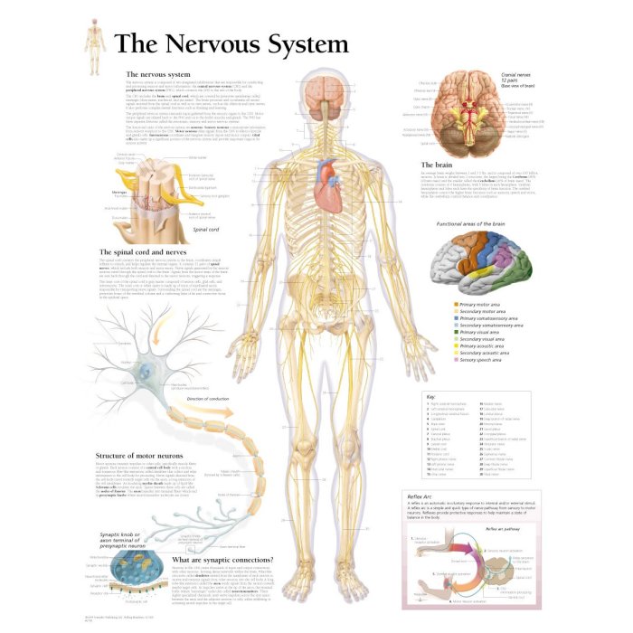
The nervous system flow chart worksheet is a comprehensive resource that provides a visual representation of the pathways and processes involved in neural communication.
How can I use the nervous system flow chart worksheet?
The worksheet can be used as a study aid, a teaching tool, or a reference guide for understanding the structure and function of the nervous system.
What are the benefits of using the nervous system flow chart worksheet?
The worksheet helps visualize and simplify complex neural pathways, making it easier to understand the intricate workings of the nervous system.